Author Archive

Rapid and easy fabrication of glass-bottom culture dishes for long-term live cell imaging

Ayako Yamada一百二十三,Jean-Louis Viovy一百二十三,Catherine Villard一百二十三和圣_潘尼除垢剂一百二十三

居里物理实验室,Institut CuriePSL Research University,CNRS UMR168,Paris,France.

Sorbonne Universités,UMPC Univ.巴黎06,Paris,法国

3皮埃尔·吉勒斯·德根尼斯研究所,Paris,法国

Email: ayako.yamada@curie.fr

Why is this useful?

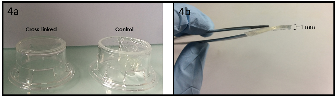
玻璃是一种多用途的化学处理表面,它仍然是迄今为止最常用的表面工程基板(例如micropatterning,surface 新利手机客户端chemistry) or plasma-bonding of PDMS microfluidic devices.在这种基质上进行细胞培养,玻璃底培养皿是为了保持细胞外明确的培养基体积,保护细胞免受污染和介质蒸发。Moreover,它们在光学上比通常用于细胞培养的聚苯乙烯培养皿更适合显微镜观察。Although glass-bottom culture dishes are commercially available (e.g.来自WPI的荧光皿)the presence of plastic walls limits the treatments that can be performed onto the glass bottom surface and those are more expensive ( 5 € per dish;φ 50 mm) than polystyrene dishes (e.g.φ 40 mm dish from TPP,0.5 € per dish).在这个提示中,we describe an easier way than a previous Tip把聚苯乙烯培养皿变成玻璃底培养皿,同时保留了在玻璃装配成盘子之前对其进行任何处理的可能性。Note that in this method,the body of the culture dish will be upside down and the lid is thus no longer lifted above the dish opening by lid stoppers.然而,the gas exchange through the gap between the body and its lid seems to be enough to culture cells healthily in this dish.In summary,this Tip provides a low-cost and rapid solution for cell culture in a microfluidic device or on an engineered surface directly in a culture dish,suited for a long-term live cell imaging.


What do I need?

  • φ 40 mm polystyrene tissue culture dish (e.g.TPP #93040)
  • φ40 mm盖板滑块(例如Thermo Fisher Scientific #11757065,每张幻灯片~0.2欧元)
  • Large screw driver (or a similar tool)
  • PDMS基和固化剂的未固化混合物(10:1 w/w)
  • 烤箱或热板
  • Ferromagnetic metal plate (e.g.PDMS容器盖,optional)
  • Cylindrical magnets (optional)


What do I do?

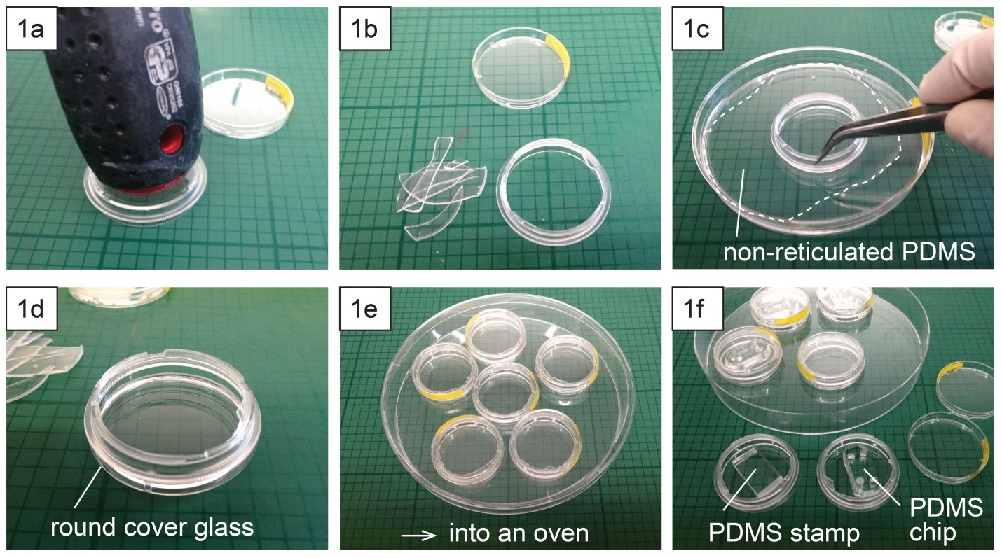
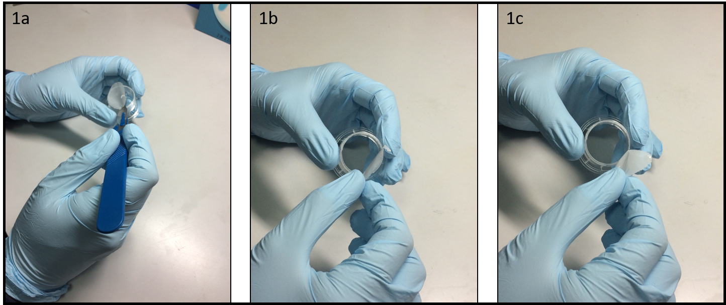
  1. Place a polystyrene culture dish upside down on a surface and hit a few times the center of the dish bottom with the grip of a large screw driver (Fig.1a) until the dish bottom falls apart from the dish wall (Fig.1b)。成功率在9%到10%之间时,底部应该容易下降。Avoid breaking the dish wall by hitting the bottom too strongly.
  2. Spread uncured PDMS mixture on a flat substrate (e.g.一个较大的塑料培养皿),并在培养皿边缘(破碎部分)涂上PDMS(图1c).
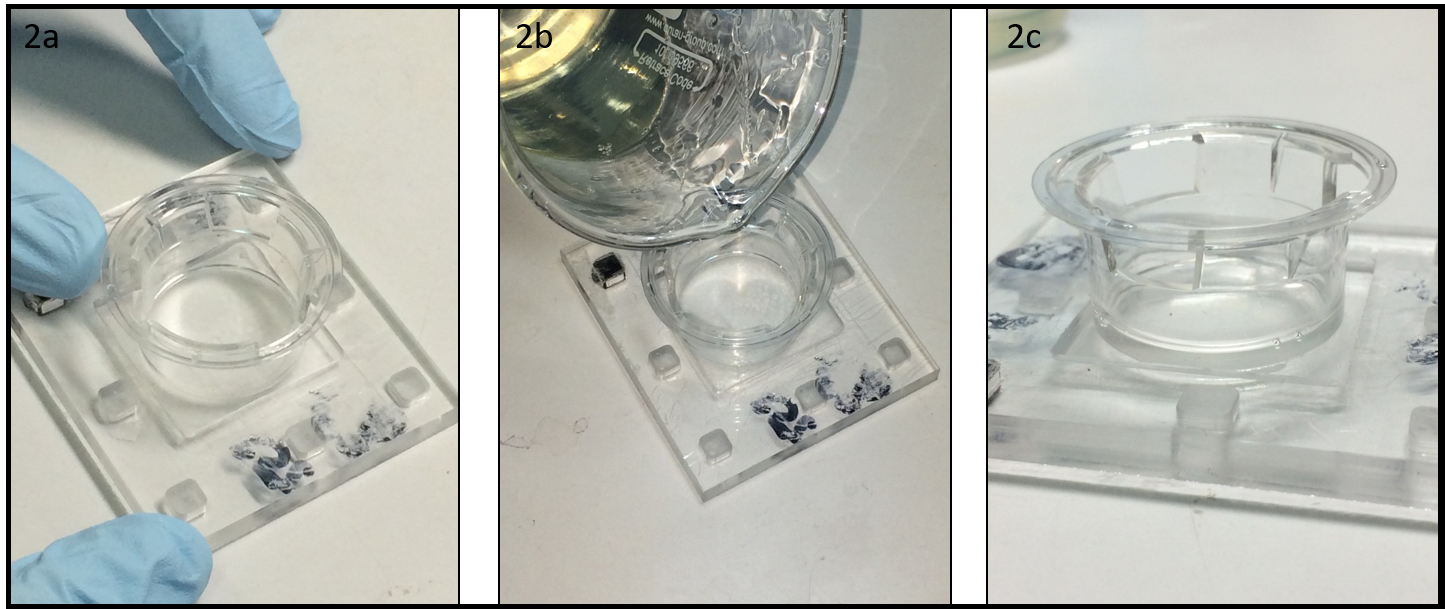
  3. Place the dish (broken part up) on a cover glass slide (Fig.1d) and cure PDMS in an oven or on a hotplate e.g.在80°C下保持10分钟(图1e).
  4. 表面处理(例如micro-contact printing) or plasma-bonding of a PDMS chip to the glass surface can be performed after or before the dish assembly (Fig.1F)。

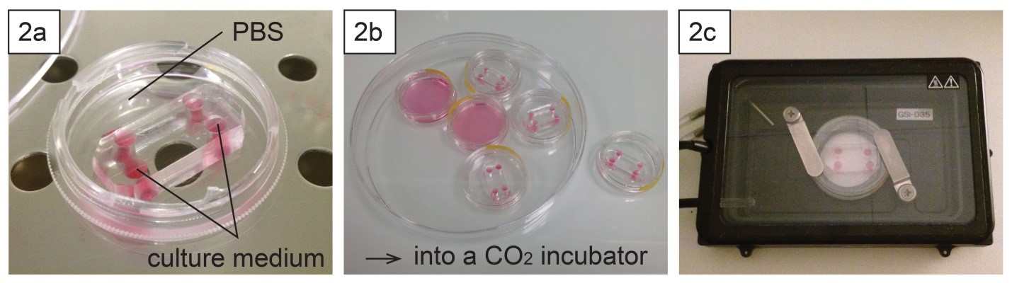
  1. 为了保持片上细胞培养的湿度,这道菜可以用例如phosphate buffered saline (Fig.2a).装有芯片或微电池的盘子可以放在一氧化碳中。有或无进一步保护的孵化器(图2b).
  2. Long-term live cell imaging can be performed using a stage top incubator (Fig.2C)。


我还应该知道什么?

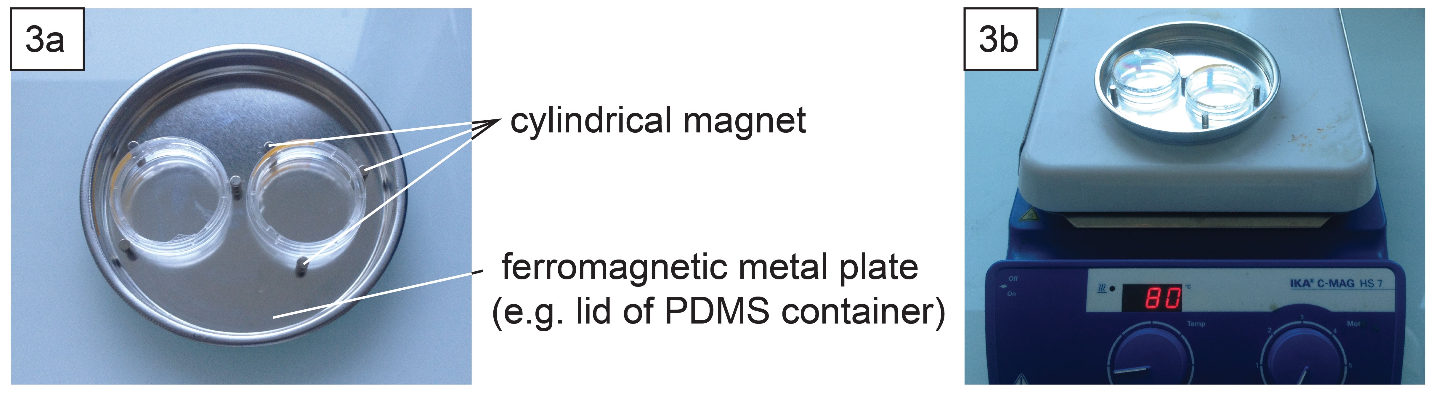
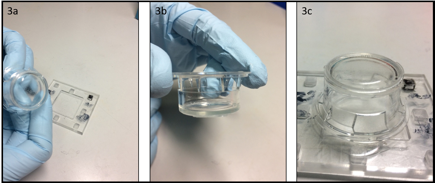
  1. 根据显微镜的支撑类型,it might be necessary to well align the contours of the dish and the glass slide.这可以使用圆柱形磁铁(每盘3个)和铁磁金属板(图3a)在烤箱或加热板上进行PDMS固化时(图3b)。


确认

这项工作得到法国国家研究机构(ANR)的支持,作为“Avenir投资计划”(参考号:ANR 10-nano 0207)和ERC高级赠款大提琴(FP7-IDEAS-ERC-321107)的一部分。

参考文献

[1] Caballero D,Samitier J为活体细胞成像研究制造细胞培养室的不同策略。Chips and Tips,2014年12月2日(//www.xcmww.com/chipsandtips/2014/12/02/different-strategies-for-the-fabring-of-cell-culture-chambers-for-live-cell-imaging-studies/)

Digg This
这就是
现在绊倒!
在Facebook上分享
Bookmark this on Delicious
Share on LinkedIn
书签在Technorati
推特上的帖子
谷歌Buzz(又名谷歌阅读器

带手动光罩对准的多层光刻

Frank Benesch Lee先生,Jose M.Lazaro GuevaraDirk R.Albrecht

Worcester Polytechnic Institute,Worcester,MA 01609 USA

为什么它有用?

现代微流控器件可以结合不同高度的通道来实现其设计功能。示例包括水动力聚焦[1],细胞陷阱[ 2 ],and chambers that isolate cellular components [3].These devices are fabricated from a multilayer SU-8 photoresist master mold.每层高度需要一套单独的光刻步骤,including photoresist spin,photomask alignment,曝光,and bakes,最后是一个开发步骤,以显示3D抵抗图案。

Mask aligners have microscopes and stage micrometers for precise,每层光罩的微米级校准,在基板晶圆上有可见的标记。它们是制作精确对准的多层图案必不可少的工具,但是在许多研究性大学的洁净室里,their substantial expense may place them out of reach of teaching institutions and individual laboratories.

相反,single-layer microfluidics can be prepared using an inexpensive UV light source,甚至是自制的。In principle,manual photomask alignment could be made under a microscope,then brought to the UV source,yet this poses several complications.第一,使用便宜的显微镜或立体镜很难看到对准特征。especially in thin SU8 layers,由于开发前暴露区和未暴露区的对比差。第二,在向曝光系统移动过程中可能会发生错位。

这里我们介绍了一种手动光罩定位方法,其精度为50微米,without the aid of a mask aligner.

What do I need?

  • Equipment and supplies for photolithography:
    • Spin coater,和紫外线曝光系统
    • 基板晶片和SU-8光刻胶
  • 小型显微镜(例如USB)或立体显微镜
  • 每层的光罩透明胶片
  • Scotch tape
  • Fine-tip permanent marker
  • Straight razor blade
  • 切割垫
  • 4 small (3/4") or mini (1/2") binder clips
  • Glass plate,大约。4×5“,与曝光系统兼容

What do I do?

  • 从透明片上切下光罩,leaving 4 corner tabs.Align the two masks relative to each other under the microscope (Figure 1a) and clip them together with a binder clip.Ensure correct mask orientation and check alignment accuracy at multiple alignment marks across the mask.(Note that horizontal alignment accuracy with a stereomicroscope is low,because each eye's optical path is angled 5 – 8 degrees,而垂直对齐不受影响。首先沿垂直方向对齐,然后将遮罩旋转90度,以确保在水平和垂直方向上精确对齐。)在每个角落添加活页夹(图1b)。并确认对准。Next,remove one binder clip at a time and use a straight razor blade to cut a sharp V-notch into each tab,through both masks.将刀片垂直向下压,以避免移动对中。Replace the binder clip,and proceed to the next corner until all 4 notches are cut (Figure 1c).

  1. 将第一层SU-8旋转至所需厚度并预烘烤。在晶圆底部贴上4片透明胶带,使粘边朝上(图2a)。Position the first mask on the wafer,pressing gently to adhere it to the tape tabs.使用细尖标记将对准缺口(图2b)追踪到透明胶带(图2c)上。转移到紫外线曝光系统并曝光。小心地移除掩模,而不将透明胶带从晶圆上拆下并烘烤后。透明胶带与95°C烘烤兼容。使用另一条胶带覆盖胶带Tabs保护标记不受污染,并允许下一个蒙版的平滑对齐。

  1. Spin coat the next photoresist layer and prebake (Figure 3a).Mount the wafer onto a glass plate with a loop of scotch tape to keep it in place.将第二个掩模放置在晶圆上,确保对齐“V”标记在每个对齐槽口内居中,并穿过所有4个角(图3b)。用薄(2-3 mm宽)胶带将面罩粘到玻璃板上,and adjust alignment as necessary.  Carefully transfer the glass plate with wafer and aligned photomask for exposure (Figure 3c).

  1. Repeat step 3 for any additional layers.取下胶带凸耳并显影光刻胶。Evaluate alignment accuracy under a microscope (Figure 4).

结论:

In this tip,我们提出了一种手动校准多个透明光罩的方法。我们实现了<100微米和50微米的可重复精度(图4a)。These accuracies are within required tolerances of many multilayer designs (Figure 4b).  In many cases,较小的设计备选方案可以放宽对准公差,such as in a trap design containing a thin horizontal channel that allows fluid bypass but captures larger objects (Figure 4c).In this example,a 100 µm wide bypass channel only partially covered the trap indentations,whereas widening the bypass channel to 400 µm enabled a functional device despite slight misalignment.  Overall,this simple method allows fabrication of microfluidic device molds containing multiple layer heights,没有昂贵的面具校准设备,to an accuracy of at least 50 µm.  Furthermore,切割对准标记后,no microscope is needed at all during the photolithography process,加速制造多个主控形状。

确认:
NSF IGERT DGE 1144804(FBL)提供的资金,Fulbright LASPAU (JMLG),University of San Carlos of Guatemala (JMLG),NSF CBET 1605679 (DRA),国家卫生研究院R01DC16058(DRA)和巴勒斯威尔康卡西(DRA)。确认:

References:

  1. Chih-Chang,C.H.Zhi-Xiong,Y.Ruey JenThree-dimensional hydrodynamic focusing in two-layer polydimethylsiloxane (PDMS) microchannels.微机械与微工程杂志,2007.17(8): p.1479.
  2. 埃里克森J.,et al.,笼状神经元MEA:一个长期研究培养的神经网络连通性的系统。神经科学方法杂志,新利手机客户端2008。一百七十五(1):P.1-16.
  3. 泰勒,a.M.,et al.,A microfluidic culture platform for CNS axonal injury,再生和运输。Nature Methods,2005.(8): p.599-605.
  4. Erickstad,M.,E.Gutierrez,and A.Groisman,A low-cost low-maintenance ultraviolet lithography light source based on light-emitting diodes.芯片实验室2015。15(1):P.57-61.

Digg This
这就是
现在绊倒!
在Facebook上分享
Bookmark this on Delicious
Share on LinkedIn
书签在Technorati
推特上的帖子
谷歌Buzz(又名谷歌阅读器

一个简单的,bubble-free cell loading technique for culturing mammalian cells on lab-on-a-chip devices

Sahl Sadeghi1­*and Meltem Elitas

工程与自然科学学院新利手机客户端Sabanci University,34956,Istanbul,Turkey

*Sahl Sadeghi写道.

目的

Lab-on-a-chip (LOC) devices significantly contribute different disciplines of 新利手机客户端science.Polydimethylsiloxane (PDMS) is one of the main materials,广泛用于制造生物基因座,due to its biocompatibility and ease of use.然而,PDMS和其他一些聚合物材料本质上是防水材料(或疏水材料)。which results in difficulties in loading and operating LOCs.在生物系统中,疏水性的显著后果是微流体通道中的气泡被截留。虽然PDMS的氧等离子体处理在一定时间内降低了表面疏水性,the hydrophilic property of PDMS vanishes over time.The persistent problem of bubbles in the microfluidics led to several studies conducted to overcome it.Some of these solutions suggested implementing bubble traps,3,surface treatment of LOCs through hydrophilic coatings4,and using actively controlled bubble removal systems,6.

尽管上述设计复杂性被引入LOC,以减少气泡引起的堵塞问题,these modifications also result in higher production cost,复杂的操作,and long device preparation time.In many single-cell experiments without losing or damaging the rare cells,这些细胞需要被导入LOC。在这里,we present a simple method that enables loading a small number of cells without introducing bubbles in the microfluidics channels.


材料

·         PDMS (Dow Corning Sylgard 184 Silicon Elastomer Kit)
·         Pipette tips (20~200μL,埃彭多夫# 3120000917)
·         Pipetman (Gilson,P200α69889-5)
·____水乙醇70% (ZAG 新利手机客户端Chemistry)
·         Cell culture medium (DMEM,PAN Biotech,#P04-01548)
·____哺乳动物细胞 (MCF7,ATCC-HTB-22)
·         Sterile syringe (bd 10毫升注射器,Luer-Lok Tip,#300912)
·____无菌汉密尔顿注射器 (汉弥尔顿,100 UL SIR,(84884)


Procedure

Step 1:Insert two 200-ul pipet tips at the inlet and outlet ports of the PDMS device as illustrated in图1.

Step 2:Introduce a 70% aqueous ethanol into the inlet-pipet tip using a pipetman.因此,the inner surface of microfluidic channels will be disinfected and the fluid flow will be tested within the micro channels as it is applied in many other protocols for LOCs,8.

Step 3:轻轻地施加压力,按压移液管,使乙醇溶液流过PDMS装置的微通道和微腔。Take care to avoid applying negative pressure from the outlet-pipet tip,which might create air leakage through the pipet connections.此外,applying a negative pressure will directly affect the amount of a gas dissolved in the liquid according to Henry's law这可能有助于在微通道内形成更多的气泡。正压有助于通过溶解气泡去除气泡。

Step 4:用乙醇溶液冲洗芯片后,检查芯片以确保去除气泡。In case of air bubbles,repeat the steps 2 and 3.

Step 5:在注射器(10毫升)中注入中等或磷酸盐缓冲盐水(PBS)。Take care to remove the air bubbles inside the syringe,安装并锁定注射器上的针头。Then,将介质流过针,确保针中充满介质,没有任何气泡。将针插入进液管尖端;轻轻地施加正压,用介质代替乙醇。Next,collect the excess medium from the outlet-pipet tip.

Step 6:Fill the inlet pipet with fresh medium in such a way that due to certain height (h) between the levels of the medium in the inlet and outlet pipet tips,微通道内将形成非常缓慢的介质流。

步骤7:Load your cells into the Hamilton syringe and take care to ensure that there is no air bubble inside its needle and syringe.如步骤5所述,将汉密尔顿注射器的针头插入进液管尖端,图1.通过向注射器施加温和的正压来引入细胞。PDMS芯片中建立的流将把释放的单元传送到芯片中所需的位置。流量可调节所施加的正压力和进出口管尖收集的介质量。The excess supernatant from the outlet-pipet tip can be collected,实验过程中可通过进口管端供给新鲜介质。


References

  1. Tan,S.H.,N.T.Nguyen,Y.C.ChuaT.G.Kang,.生物流体学,2010.4(3).
  2. Zheng,W.F.Z.王W张and X.Y.Jiang,. 芯片实验室,2010.10(21): p.2906-2910.
  3. 王Y.d.Lee,洛杉矶张H.Jeon,J.E.Mendoza EliasT.A.哈瓦特S.Z.Hassan,a.Zhou,D.T.Eddington,and J.Oberholzer. Biomedical Microdevices,2012。十四(2):P.419-426。
  4. 王Y.L.,C.E.模拟人生and N.L.Allbritton. 芯片实验室,2012。十二(17): p.3036-3039。
  5. 卡尔森J.M.M.盖曾S.Laakso,THaraldsson,S.Malhotra-Kumar,M.Maki,H.Goossens,and W.范德威格纳特,. 芯片实验室,2013。13(22): p.4366~437
  6. Cortes,D.F.T.-X.唐D.G.S.Capelluto,I.M.Lazar,. Sensors and Actuators B: Chemical,2017。243P.650-67.
  7. Benavente-Babace,A.,d.Gallego PerezD.J.HansfordS.AranaE.Perez Lorenzoand M.穆吉卡. Biosensors & Bioelectronics,2014。61P.98-305。
  8. Yesilkoy,F.,R.Ueno,B.X.E.Desbiolles,M.Grisi是的。Sakai,B.J基姆,and J.布鲁格. 生物流体学,2016。10(1).
  9. Henry,W.,Phil.反式R.Soc.Lond.,1803。九十三:29—274。

Figure 1 – Schematic of cell loading procedure in a microfluidic PDMS device.

Digg This
这就是
现在绊倒!
在Facebook上分享
Bookmark this on Delicious
Share on LinkedIn
书签在Technorati
推特上的帖子
谷歌Buzz(又名谷歌阅读器

一种新的低成本制备交联明胶膜的方法

Gabriele Pitingoloand Valerie Taly

INSERM UMRS1147,CNRS SNC 5014,巴黎笛卡尔大学Equipe labellisée Ligue Nationale contre le cancer 2016.Paris,France.

email:gabriele.pitingolo@parisdescartes.fr

Why is this useful?

近年来,几种明胶类型(即GelMA,A or B) have been used in pharmaceutical formulation and tissue engineering due to their excellent biocompatibility,propensity to cell differentiation and availability at low cost.The use of gelatin as biomaterial is also advantageous for the possibility to tune the mechanical properties of the substrate,changing the concentration in water and the degree of cross-linking.然而,Transwell®is the most used permeable support with microporous membranes and is a standard method for culturing cells.This commercial type of support has been widely used to study the molecular secretion by different cell types and also to reproduce severalin vitrophysiological barriers (i.e.血脑屏障)。3Transwell®细胞培养插入很方便,because they are sterile and easy-to-use,but they are very expensive (~ 300 $ for a 12 pack) and possess a limited range of biomaterial properties because they are made from polyester or polycarbonate.此外,as shown by Falanga and colleagues,这些多孔膜通常集成在微流控芯片上进行渗透性研究。4

Recently,Yong X.陈等人。proposed an alternative method to prepare a suspended hydrogel membrane platform for cell culture,设计了一个复杂的合成凝胶的协议,并用商用打印机制作了一个由聚乳酸(PLA)聚合物制成的开放式网格结构。Our tip shows a novel low-cost method for preparing a cross-liked gelatin membrane as a permeable support useful for potential biological applications.In addition,提议的方案不需要使用复杂的制造技术或昂贵的材料。它使用来自猪皮肤的明胶和甲醛蒸气技术交联明胶膜。作为概念证明,we integrate the gelatin membrane into a microfluidic chip,to show the possibility to develop a platform for comparable studies in static and dynamic conditions.

此外,为了证明制备的明胶膜对培养温度(约37°C)的抗性,我们在孵育前后(2天)测试了力学性能(杨氏模量)。Finally,我们观察了机械性能和结构完整性的保存,使膜可用于细胞培养的研究。


What do I need?

  • Transwell插件
  • Porcine gelatin type A
  • 甲醛溶液
  • 手术刀
  • PMMA研磨室或类似

What do I do?

  1. 从Transwell中取出多孔膜®insert (Fig.1a-1c) or alternatively use a similar homemade support.为了方便这一步是方便使用解剖刀沿整个直径切开膜。

  1. 膜移除后,Transwell®支持随时可用。Position the structure at the center of the PMMA chamber (depth 2 mm at least) (Fig 2a) and pour liquid 10% w/v gelatin,without bubbles,onto the PMMA chamber and inside the Transwell®support (Fig 2b).稳定2分钟后,put the system in the fridge,for at least 10 minutes.

  1. After the gelation time (10 minutes at 4°C) it is possible to remove the formed gelatin membrane from the PMMA chamber,借助手术刀促进分离(图3a-3b)。如图3c所示,明胶膜看起来非常平坦,an ideal characteristic for cell cultivation (Fig 3c).To guarantee the preservation of the mechanical properties during the cell culture step,cross-link the gelatin membrane using the classical protocol "cells-biocompatible",such as glyceraldehyde6,甲醛and glutaraldehyde8methods or natural products such as genipin.

  1. In this case,采用气相甲醛法将制备的明胶膜交联,得到水溶性较低的体系。higher mechanical strength and stability against enzymatic degradation.我们将明胶膜暴露在甲醛蒸气中1天。在图4a中,我们显示了差异,培养48小时后,between a sample cross-linked (left) and not (right).交联明胶膜的最终深度约为1 mm,然而,it is possible to change the depth tuning the liquid gelatin amount.Finally,我们计算,before and after the incubation time,交联明胶膜的杨氏模量,observing a similar value of 40 kPa (compression test by using hydraulic testing system Instron DX).

  1. Integration of the gelatin membrane into a microchip.In this section,we detail the integration of the gelatin membrane into a microfluidic chip.As example,we used the same geometry proposed in our previous work4,to make a device for a permeability experiment (Figure 5a).如图5b所示,just pour the liquid gelatin into the smaller drilled microchannel,using a pipette to form a thin uniform layer of gelatin (Figure 5c).After gelation at 4° C,cross-link the formed gelatin membrane using the previously described method,结果如图5d所示。为了连接不同的PMMA-PDMS基板,我们在这里提出了一种我们小组最近开发的磁性方法。10,通过物理化学应力保护明胶膜,如溶剂蒸发和等离子粘合。Figure 5e shows the final chip.


结论:In this tip,a novel biocompatible gelatin permeable support was obtained by using a simple and low cost fabrication method.可采用气相甲醛法或其他化学交联方法对整体明胶膜进行交联,作为细胞培养的潜在支架。此外,通过改变明胶的初始浓度,可以调节渗透膜的杨氏模量和厚度。the degree of cross linking and the amount of liquid gelatin.Finally,we showed the integration of the gelatin membrane into a modular microchip.因此,we propose an easy and low cost method to prepare a permeable gelatin membrane for cell biology and for other applications.

确认

This work was carried out with the support of the Pierre-Gilles de Gennes Institute equipment ("Investissements d'Avenir" program,reference: ANR 10-NANO 0207).

References

  1. Geckil,Hikmet,et al.“作为细胞外基质模拟的工程水凝胶”,纳米医学5.3(2010):469-484。
  2. https://www.corning.com/worldwide/en/products/life-新利手机客户端sciences/products/pervative-supports/transwell-guidelines.html
  3. GuarnieriDaniela,et al."Shuttle‐Mediated Nanoparticle Delivery to the Blood–Brain Barrier." Small 9.6 (2013): 853-862.
  4. Falangaa.P.,Pitingolo,G.Celentano,M.,CosentinoA.,梅隆P.,VecchioneR.NETTI,P.a.(2016)。在流动条件下穿梭介导的纳米颗粒在体外大脑内皮的转运。Biotechnology and Bioengineering.
  5. Chen,Yong X.et al.“用于细胞培养的新型悬浮水凝胶膜平台”,《工程与医学纳米技术杂志》6.2(2015):021002。
  6. Sisson,Kristin,et al."Evaluation of cross-linking methods for electrospun gelatin on cell growth and viability." Biomacromolecules 10.7 (2009): 1675-1680.
  7. 美国宇航局M.,et al.“整齐填充明胶的行为和特性”,《生物材料》24.1(2003):165-172。
  8. Talebian,A.,et al."The effect of glutaraldehyde on the properties of gelatin films." Kemija u industriji 56.11 (2007): 537-541.
  9. BigiA.,et al."Stabilization of gelatin films by crosslinking with genipin." Biomaterials 23.24 (2002): 4827-4832.
  10. 皮提戈洛·加布里埃尔,et al."Fabrication of a modular hybrid chip to mimic endothelial-lined microvessels in flow conditions." Journal of Micromechanics and Microengineering" (Accepted manuscript)
Digg This
这就是
现在绊倒!
在Facebook上分享
Bookmark this on Delicious
Share on LinkedIn
书签在Technorati
推特上的帖子
谷歌Buzz(又名谷歌阅读器

DIY蠕动泵

Shannon FaleyBradly Baer马修·理查森,Taylor Larsen,Leon M.贝兰*

范德比尔特大学Department of Mechanical Engineering,Nashville TN,37235,USA

*leon.bellan@vanderbilt.edu

Why is this useful?

Figure 1: Fully assembled peristaltic pump


The majority of microfluidic applications require an external pumping mechanism.  Multi-channel,individually addressable pumps are expensive,often large,and prone to failure when operated inside cell culture incubators at 95% humidity.  The number of experiments that can be run at a given time is limited by the availability and expense of pumps.  Perfusing artificial tissue scaffolds containing engineered vasculature requires long-term (days to weeks) continuous flow at low rates.  We designed an inexpensive (~$100 for 2 pumps,~$70 for each additional set of 2 pumps) peristaltic pumping system using an Arduino- controlled stepper motor fitted with a custom 3D-printed pump head and laser-cut mounting bracket.Each pump has a footprint roughly that of the NEMA 17 stepper motor and is easily controlled individually using open source software.  Up to 64 motor shields can be stacked for a given Arduino Uno R3,each capable of supporting two stepper motors,这样就有了扩展潜力,可以同时控制128台泵。我们成功地实现了两个堆叠的电机护罩,驱动四个独立的步进电机。流速取决于管子直径和步进速度。我们发现1/16“管子的流速范围在~50-250μl/min之间,1/4”管子的流速范围在~500-1500μl/min之间。我们预计,与直流电机驱动的标准蠕动泵相比,这种泵的设计可能对培养箱湿度更有弹性。自从实施离子,这些泵在潮湿条件下连续运行3个月(间歇)。In the event of failure,然而,cost of motor replacement is an economical $14.

Figure 2

我需要什么?


材料:

  • NEMA 17步进电机(14美元,spec,vendor)
  • Arduino UNO R3控制器(25美元,spec,vendor)
  • Arduino Motor Shield(20美元,spec,vendor)
  • M3机械螺钉(4)和六角螺栓(4)($1,McMaster-Carr)
  • DB9 Male & Female Solder Connectors ($9,星际科技
  • 18AWG 4C扬声器电缆(10美元,Monoprice)
  • 弹簧钢
  • ABS Filament
  • 6-32机械螺钉和方螺母(3)($1,McMaster-Carr)

Equipment:

  • 3D打印机
  • Laser/Metal cutter
  • Soldering iron & solder
  • 丁烷焊炬

我做什么?


Pump head fabrication:

  1. Using ABS filament,文件中的3D打印泵头pumphead.crt.9
  2. 从刚性的1/4”管子上切下三个15 mm(长)的部分,用作滚轴。
  3. Use the three 6-32 machine screws and square nuts to assemble the tubing to pump head as shown in Figure 3.

图3

Mounting bracket fabrication:

  1. Using bracket template file(2000泵安装v4)以及激光切割设备,用弹簧钢制作安装支架,or other appropriate metal.  Note that the score line bisecting the bracket is intended to be cut at a lower power.  This line is just a marker to show where to bend the bracket in the following step.
  2. 使用手持丁烷火炬,沿划线加热安装支架并用钳子弯曲。重复此操作,直到安装支架形成直角(见图1)。

Motor Electrical Wiring:(方向示例见图4)

  1. Solder motor wires to DB9 Male Connector
  2. Solder one end of speaker wire to DB9 Female Connector
  3. Connect opposite end of speaker wire to Arduino Motor shield

Figure 4 Example of connection scheme by wire color

Pump Assembly:

  1. Use M3 machine screws to attach mounting bracket to stepper motor,with corresponding hex nuts as spacers between motor and bracket.
  2. Press fit pump head onto rotor shaft.
  3. 使用DB9连接器将电机连接到Arduino

Arduino/电机屏蔽组件:

  1. 遵循adafruit.com提供的组装说明(https://learn.adafruit.com/adafruit-motor-shield-v2-for-arduino/stacking-shields).另见图5。

Figure 5

计算机控制:

1.请参阅联机资源以获取简单的入门代码https://learn.adafruit.com/adafruit-motor-shield-v2-for-arduino/install-software)

2.Load example code to control 2 stacked motor shields running four independent pumps simultaneously.(四步进电机

三。Start pumping!  See  video clip for multi-pump demonstration:


Please click here to download the DIY Peristaltic Pump Files

Digg This
这就是
现在绊倒!
在Facebook上分享
Bookmark this on Delicious
Share on LinkedIn
书签在Technorati
推特上的帖子
谷歌Buzz(又名谷歌阅读器