Archive for the ‘Hot Articles’ Category

Why should we use optofluidics for monitoring marine environment?

Phosphorus is found in natural waters and exerts a major influence on the composition and structure of aquatic ecosystems. It is a crucial nutrient for planktons and algae, which feed fish and other marine organisms. However, human activities may result in excess amount of phosphorus, which, in turn, causesharmful algaeto bloom in natural waters. The bloomcreates a hostile environmentto other forms of marine life by consuming the available oxygen in the sea, and producing toxins. Sea organisms such as fish swim away from the blooms, but the ones that cannot swim, such as shellfish, unfortunately die. We do care about this occurrence as it negatively affects natural life and the economy. There is only one way to interpret the effect of continuously changing phosphorus levels on the strength of the biological pump: real-time monitoring of phosphate levels in the marine environment!

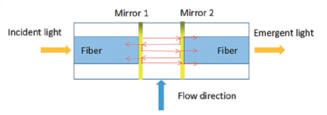
Figure 1. The design of the Fabry-Pérot microcavity, consisting of two parallel mirrors (reflectors) fabricated by coating the surface of the optical fibers with a gold layer. The light is reflected by the mirrors multiple times to enhance the signal. Adapted from Zhu et al., 2017.

Conventional vs. optofluidic monitoring instruments

Conventional phosphate monitoring instruments are mostly used for on-site sampling, then the fresh sample is transported to a laboratory for determining the phosphate level. Laboratories complete one round of analysis in 20 min, often using spectrophotometrical measurement tools. Given the conditions, real-time phosphate monitoring easily becomes laborious, time consuming, and costly. To address this challenge, researchers in Chinese Academy of Sciences, Wuhan University, and The First Institute of Oceanography in China collaborated to develop a portable optofluidic phosphate monitoring tool. However, prototyping an optofluidic marine phosphate detection tool is not straightforward because anabsorption cell—a component core to the measurement unit—is simply too big to fit in a microchip. Instead of using a bulky absorption cell, researchers considered integrating aFabry-Pérot cavityin the microsystem. The Fabry-Pérot cavity consists of two parallel optical fibers with a spacing in between. The cross-sectional surface of each optical fiber is coated with a thin layer of gold to create reflector surfaces (Figure 1) in order to enhance the absorption of phosphate. Shortening the spacing between the reflectors decreases the analysis time from minutes to seconds.

How it works?

In the microchip, filtered water sample and a chromogenic reagent are injected into a curved microchannel. After the chromogenic reaction, the water-soluble components are transported into the optical section (Figure 2). The probe light is sent into the Fabry-Pérot cavity via one of the fibers, bounces between the reflectors multiple times to increase the optical feedback and then analyzed by the detector. The obtained absorbance value, therefore, increases linearly with increasing phosphate concentration. In this microsystem, phosphate detection range is 0.1-100 µmol per liter (400 times greater than the range of a conventional instrument) and detection time is 4 seconds (300 times shorter than detection time of a conventional instrument). The authors of the paper think that this technology can be applied to detect other nutrient levels as well as pH changes in marine environment.

optofluidic phosphate monitoring

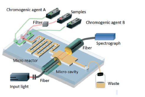
Figure 2. A schematic of the optofluidic microchip consisting of two parts: the microfluidics circuit forming the microreactor in the microchannel, and the optical part to provide optical feedback for enhanced absorption analysis. Adapted from Zhu et al., 2017.

To download thefull article for free*click the link below:

Optofluidic marine phosphate detection with enhanced absorption using a Fabry–Pérot resonator

M. Zhu, Y. Shi, X. Q. Zhu, Y. Yang, F. H. Jiang, C. J. Sun, W. H. Zhaoc, and X. T. Hanc

Lab Chip, 2017, Lab on a Chip Recent Hot Articles

DOI:10.1039/C7LC01016H

About the Webwriter

Burcu Gumuscuis a postdoctoral fellow inHerr Labat UC Berkeley in the United States. Her research interests include development of microfluidic devices for quantitative analysis of proteins from single-cells, next generation sequencing, compartmentalized organ-on-chip studies, and desalination of water on the microscale.

*until 16th February 2018

Digg This
Reddit This
Stumble Now!
Share on Facebook
Bookmark this on Delicious
Share on LinkedIn
Bookmark this on Technorati
Post on Twitter
Google Buzz (aka. Google Reader)

Spotting cancer: one in a billion cells

The human body consists of tens of trillions cells, all of which theoretically should have the same genome. Depending on genetic and environmental factors, some of these cells experience point mutations. Although most of those mutations are cleaned up by DNA repair enzymes, about 0.01% of them stay. A low percentage of the persistent mutations turn out to be ‘cancer’ while others stay recessive. Several genes, including the ones responsible for cell growth cycle, cause the persistent mutations. Uncontrolled growth of cells leads to formation of tumor, which is now prone to experience more mutations due to continuous proliferation. These mutations create heterogeneity among the cell population of a tumor, and eventually some cells leave their original tumor and start a new one in another organ of the body. When a cell leaves its parent tumor, it starts circulating in blood vessels before settling in. Those cells are calledcirculating tumor cells(CTCs), and around1-10 CTCcan be found in 1 mL of blood (which contains about1 billion red blood cells, and 1 million white blood cells). Capturing ultra-rare CTCs has enormous implications in early cancer diagnosis. Often times, analysis of at least 10 mL of blood is necessary to capture sufficient CTCs to confirm their presence. The available technology can achieve CTC capturing in about 10 hours leading to the loss of target cells and decay of detection biomarkers.

Microfluidic devices are well known for precise sorting of microscale materials. Parallelizing microscale sorting in microfluidic devices enables high-throughput sample processing. In the light of this principle,David Issadore, Jina Ko, and their fellow researchers at University of Pennsylvania coinedCaTCh FISH, a circulating tumor cell fluorescence in situ hybridization platform for rapid detection of CTCs. David Issadore kindly accepted to talk about this exciting lab-on-a-chip device. According to David, “the main strength of the CaTCh FISH is that it preserves the sensitivity and specificity of microfluidic cell sorting and RNA FISH, but through clever engineering allows these normally very slow laboratory-based operations to be performed rapidly and automatically on a chip.”

RNA FISH, high-throughput cell handling

Figure 1. Overview of the CaTCh FISH platform. Whole blood sample is processed in TEMPO step, where magnetic nano particle based cell separation is followed by single-cell RNA analysis (modified from Ko et al., 2017).

Processing a whole blood sample using CaTCh FISH involvesthree steps(Figure 1). First, white blood cells are labelled with magnetic nanoparticles. Second, whole blood is passed through a magnetic micropore filter to selectively trap magnetically labelled cells. “Our magnetic micropore device rapidly and precisely removes all of the cells that we know are not CTCs”, says David. The operating principle of magnetic micropore filter is based on strong and highly localized microscale field gradients formed at the edge of micropores to enable application of high flow rates. Third, single cell RNA analysis is performed on isolated cells using rapid in-situ hybridization strategy so that CTCs can be identified within the isolated cell population. In this way, targeted CTCs can be isolated from the rest of the cell population regardless of their physical and molecular properties. Analysis of a 10 mL blood sample takes less than an hour.

As akey novelty, the researchers maintainedhigh-throughput processing and high sensitivity at the same timeby integrating the FISH technique (hybridization of 20-50 fluorescently-labelled oligonucleotide probes to the target RNA, and subsequent fluorescence-signal based detection to enhance signal-to-noise ratio) in a microfluidic chip. CaTCh FISH has also beentested in patients with pancreatic cancerand detected CTCs in the real patient samples.

“The CaTCH FISH technology can be easily modified to measure other rare cells, for the diagnosis of other cancers or for stem cell research for example, by modifying the RNA FISH probes”, says David. He considers converting the platform to a high-precision hospital-based diagnostic tool and he collaborates with a company in the Bay area for this process. The CaTCh FISH device is poised to have a big impact on the way cancer is diagnosed.

To download thefull article for free*click the link below:

A magnetic micropore chip for rapid (<1 hour) unbiased circulating tumor cell isolation and in situ RNA analysis

Jina Ko, Neha Bhagwat, Stephanie S. Yee, Taylor Black, Colleen Redlinger, Janae Romeo, Mark O’Hara, Arjun Raj, Erica L. Carpenter, Ben Z. Stanger and David Issadore

Lab Chip, 2017, Paper

DOI:10.1039/C7LC00703E

About the Webwriter

Burcu Gumuscuis a postdoctoral fellow inHerr Labat UC Berkeley in the United States. Her research interests include development of microfluidic devices for quantitative analysis of proteins from single-cells, next generation sequencing, compartmentalized organ-on-chip studies, and desalination of water on the microscale.

*until 5th January 2018

Digg This
Reddit This
Stumble Now!
Share on Facebook
Bookmark this on Delicious
Share on LinkedIn
Bookmark this on Technorati
Post on Twitter
Google Buzz (aka. Google Reader)

Zenith in “artery”

When cutting a finger, thrombocytes and fibrin in the blood make up the blood-clotting mechanism,aka.haemostasis, to stop the blood loss. Another way to trigger this mechanism is having an artery damaged byatherosclerosis,这通常是由多个基因或acqu引起的ired factors. In the latter case, thrombosis develops within a vein or artery, obstructing or stopping the blood flow to major organs like the heart and eventually causing heart attack. Considering every year over 14 million lives worldwide are lost to heart attacks, more investigation on this topic is needed without any doubt.

Recently, a research team led byAndries van der Meerpublished a research article inLab on a Chipon mimicking arterial thrombosis in 3D vascular structures, representing a major step forward in the development of accurate and faster methods of studying arterial thrombosis without using animals. The authors highlighted the inconvenience of using animal models to predict arterial thrombosis in humans. This is mainly due to fundamental differences between human and animal physiology, the researchers explain. For instance, rodent platelet biology, coagulation dynamics, and shear stress in mice arteries significantly vary between humans and mice.

thrombosis on chip

Figure 1. Three-dimensional models of a healthy and stenotic vessels and thrombosis formation upon blood perfusion through the channels.

The paper uses miniaturized vascular structures mimicking 3D architectures found in both healthy and stenotic blood vesselsin-vitro(Figure 1). They combinedstereolithographyand 3D printing of computed tomography angiography data to construct 3D-printed templates of vessels inPDMSmicrochips. The 3D printed vessels are then coated withhuman umbilical vein endothelial cells, forming a monolayer fully covering the surface. In the next step, the artificial vessels are perfused with blood at normal arterial shear rates, allowing a blood clot to form as it would happen in the human body. The 3D printed vessel is clinically more relevant when compared to 2D vessel models, since the realistic flow profiles of blood and even distribution of shear stress across the vessel are of great importance when researching arterial thrombosis. Hugo Albers, the co-first author of the paper explains what led the team to try 3D models:“Other groups have worked on thrombosis-on-a-chip before, but we wanted to incorporate flow profiles that are similar to what one would find in-vivo. So we opt for a round and thus 3D shape. Since the stenotic geometry is an important part of this work, we wanted to find a technique that allowed us to make almost any shape we could come up with. Thus 3D-printing seemed to be the way to go.”

When it comes to defining the challenges in 3D organ-on-chip modeling and fabrication, “我们需要复制的细胞环境ing human endothelial cells and human whole blood to fully mimic the nature of vasculature” says Albers. “Incorporating the shape of vasculature to recreate the flow profiles found in-vivo and recreating the shape of vasculature on a small scale was quite challenging, since the resolution of 3D-printing quickly started to be the limiting factor. Furthermore, we ran into problems related to working with whole blood. We had to figure out how to perfuse small channels with blood without instigating thrombosis outside of the microfluidic channel.” The researchers successfully overcame the challenges mentioned by Albers and mimicked the formation of thrombosis in a stenotic vessel model as seen in Figure 1 (bottom).

The researchers note that the next step involves co-culturing arterial endothelial cells and smooth muscle cells with human umbilical vein endothelial cells or moving to different cell lines such as differentiated human induced pluripotent stem cells. “I think we can also apply the 3D-printing technique to create thrombosis-on-a-chip devices with different geometries, e.g. aneurysms or bifurcated geometries”, says Albers.

To download thefull article for free*click the link below:

Mimicking arterial thrombosis in a 3D-printed microfluidic in vitro vascular model based on computed tomography angiography data

Pedro F. Costa, Hugo J. Albers, John E. A. Linssen, Heleen H. T. Middelkamp, Linda van der Hout, Robert Passier, Albert van den Berg, Jos Malda and Andries D. van der Meer

Lab Chip, 2017, Paper

DOI:10.1039/C7LC00202E

This paper is included in our Organ-, Body- and Disease-on-a-Chip Thematic Collection. To read other articles in the collection, visit –rsc.li/organonachip

About the Webwriter

Burcu Gumuscuis a postdoctoral fellow inHerr Labat UC Berkeley in the United States. Her research interests include development of microfluidic devices for quantitative analysis of proteins from single-cells, next generation sequencing, compartmentalized organ-on-chip studies, and desalination of water on the microscale.

Digg This
Reddit This
Stumble Now!
Share on Facebook
Bookmark this on Delicious
Share on LinkedIn
Bookmark this on Technorati
Post on Twitter
Google Buzz (aka. Google Reader)

Droplets droplets everywhere: optimising genetic circuits using cell-free extracts and droplet microfluidics

The design and engineering of biological systems — a field known asSynthetic Biology—has been heralded by many to be the defining technology of the 21stcentury. Just as electronics and computers have changed almost every facet of our day-to-day lives, the consequences ofengineering microbesmight also be as far reaching. Engineered bugs are already being developed to produce pharmaceuticals and fuels, monitor their environment, detect trace chemicals, degrade plastic waste, and perform computing operations.

Microbes, however, are considerably more complex — and hence less predictable — than electronic components. For this reason, one of the best strategies scientists have for engineering them is to use a brute force approach: try out as many different biodesigns as possible and see what works best.

Doing this with microbes is tricky. Designing and constructing a single engineered bacteria is labour intensive and can take weeks. To circumvent this, researchers have turned tocell-free systems: a collection of biomolecules that can reproduce biochemical features of interest, for example the synthesis of proteins from a genetic circuit. This greatly simplifies things as after all you are now merely handling what is essentially a sack of chemicals.

This is where it really gets interesting. As opposed to testing out different genetic circuits one-by-one in a test tube,Horiet aldeveloped a microfluidic device which forms millions of droplets an hour – each droplet acts as a mini test tube, each containing a slightly different version of the circuit. The circuit was made up of three strands of DNA which interact with one another in a non-linear way to produce a Green Fluorescent Protein. The fact that it is non-linear means predicting how they interact is difficult and makes the tactic of trying out different combinations more appealing. Using microfluidics, the authors created a generic library by combining different ratios of these three components. By incubating the droplets and monitoring protein production levels, they were able to map out the parameter space to see which genetic circuit from their library worked best.

Although simply a proof-of-concept, this droplet microfluidic approach has shown to be incredibly powerful, and in addition to being used ingenetic circuit designhas the potential to rapidly explore large parameter spaces in other areas of chemistry and biology.

To download thefull article for free*click the link below:

Cell-free extract based optimization of biomolecular circuits with droplet microfluidics
Yutaka Hori, Chaitanya Kantak, Richard M. Murray and Adam R. Abate
Lab Chip, 2017
DOI: 10.1039/C7LC00552K

*Free to access until 8th September 2017.


About the Webwriter

Yuval Elaniis an EPSRC Research Fellow in the Department of Chemistry at Imperial College London. His research involves using microfluidics in bottom-up synthetic biology for biomembrane construction, artificial cell assembly, and for exploring bionterfaces between living and synthetic microsystems.

Digg This
Reddit This
Stumble Now!
Share on Facebook
Bookmark this on Delicious
Share on LinkedIn
Bookmark this on Technorati
Post on Twitter
Google Buzz (aka. Google Reader)

3D Printing Truly Micro Microchannels

A new 3D printer and resin formula enable 3D printing of channels on the microscale

There is no doubt that 3D printing is a hot technology with applications across all sectors of industry and life. The technology is attractive not only for its use in rapid prototyping, but also for its ability to form complex structures that could not easily be formed by other manufacturing techniques. There is also the added benefit of digital design—it is very easy to go from a CAD model to physical production without the need of additional tooling or equipment. For these reasons and others,3D printing is an attractive manufacturing process for microfluidics.

Albert Folch has argued that 3D printing is the solution to the barriers preventing the translation of PDMS microfluidics to commercial applications. (Angew. Chem. Int. Ed.2016, 55, 3862–3881.) However, printing resolution haslimited the size of channels to the milli- and sub-millifluidic regimes. The challenge when it comes to microchannels is that commercial 3D printers are designed to form polymer features but microchannels, by their nature, arevoidswithin bulk material. While many 3D printer manufacturers tout resolutions in the tens of micrometers, the reality is that the resolution of the void spaces can be several-fold larger. In theirrecent report, researchers at Brigham Young University have advanced 3D printing technology to print channels truly in the micro- regime.

Left: Schematic illustration of 3D stacked serpentine channel design. Right: SEM image of 3D serpentine channel cross section.

Achieving true micro-scale printing resolution required innovation in both the 3D printer and the resin material. Gonget al.modified a digital light processing (DLP)-type 3D printer to include a high resolution light engine and a 385 nm UV LED light source. DLP printers project a pattern of light over the entire plane, crosslinking an entire layer in a single step. The light engine enables resolutions in thex-yplane of 7.6 µm and their choice of a 385 nm light source opened a wide range of possible UV-absorbers to them. Twenty different UV-absorbers were evaluated and 2-nitrophenyl phenyl sulfide (NPS) was chosen as having all the desired properties, with the main criterion of enabling smaller void sizes in thez-dimension. The final composition of the resin was a mixture of poly(ethylene glycol) diacrylate (PEGDA), NPS, and Irgacure 819. This mixture created structures that have low non-specific adsorption and had good resolution in thez-axis. Additionally, the structures could be photocured with a broad-spectrum UV lamp after printing.

Despite high-resolution printers and materials, a new printing regime also had to be developed to achieve the desired channel width. Because of scattering, the area of crosslinked resin tended to be smaller than the actual pixel size. This meant that channels, which are essentially voids of non-exposed space, came out wider than designed. The solution that Gonget al.devised was to perform two print patterns for each exposed layer. The first print pattern exposes the entire area, save for the channel void and the second exposure only projects a pattern along the channel walls. This method enabled reproducible printing of channels with a ~20 µm width.

3D CAD model of the custom 3D printer by Gong et al.

So how soon can you expect to get your hands on a 3D printer capable of printing true microchannels? While this report has been very promising, it is currently the only printer and only resin formulation with these capabilities. According to Greg Nordin, the corresponding author, the main challenge is to figure out “how to get this into people’s hands in a convenient way so they don’t have to mess around much to get it to work.” Like any new technology, for it to be truly useful, users want to spend more time making and less time fiddling. Fortunately, there already are markets for printing small, intricate parts, such as jewelry and dentistry, so it’s only a matter of time before manufacturers catch on to the market for 3D printed microfluidics.

To download thefull article for free*click the link below:

Custom 3D printer and resin for 18 μm × 20 μm microfluidic flow channels
Hua Gong, Bryce P. Bickham, Adam T. Woolley and Gregory P. Nordin
Lab Chip, 2017
DOI: 10.1039/C7LC00644F

*Free to access until 29th August 2017.


About the Webwriter

大流士Rackus博士后研究员University of Toronto working in theWheeler Lab. His research interests are in combining sensors with digital microfluidics for healthcare applications.”

Digg This
Reddit This
Stumble Now!
Share on Facebook
Bookmark this on Delicious
Share on LinkedIn
Bookmark this on Technorati
Post on Twitter
Google Buzz (aka. Google Reader)

How can biosensors provide real-time health monitoring?

Like a ripple spreading outwards on a pond’s surface, aplasmonis a collective oscillation of free electrons in a piece of metal. When the metal interacts with light (in this casek vector光满足动力条件),有限公司llective movement of oscillating electrons at the metal’s surface leads to the propagation of the light along the surface, also known as a surface plasmon. This simple physics rule is very helpful for sensor nanotechnology and optical signal processing applications since the offered advantages are significant, providing a smaller foot-print, lower limits of detection, and multiplexing opportunities. However, using sensors for biomolecule detection requires clever use of light. For example, surface plasmon resonance can be observed in nanohole arrays patterned on very thin gold films. Nanohole arrays can transmit light much more strongly than expected for the nanohole apertures at certain wavelengths. This phenomenon is calledextraordinary optical transmission(EOT) and opens a new avenue for detection of minute amounts of biomolecules.

A nanoplasmonic biosensor was recently developed for real-time monitoring of proteins from live cells in a label-free configuration, thanks to extraordinary optical transmission. The biosensor is structured in a microfluidic device consisting of a cell module and the biosensor module. Microfluidic cell module design is based on a single zigzag channel for directly delivering the secreted proteins to the adjacent biosensor module via a tubing connection. The biosensor module consists of nanoholes fabricated on freestanding SiNx membranes. Antibodies specific tovascular endothelial growth factor(VEGF) are selectively immobilized on the gold nanohole arrays as biorecognition molecules. When the VEGF are captured on the sensor, a change of refractive index proximate to the surface is induced. This change results in a peak shift of the EOT spectrum (Figure 1). By tracing the spectral shifts continuously, the dynamics of cell secretion can be monitored. In this way, the secretion dynamics from live cancer cells are monitored and quantified for 10 hours. The proposed microfluidic device has unique capabilities for multiplexed and label-free detection in a compact footprint, which are promising for miniaturization and integration into lab-on-a-chip devices.

biosensor, microfluidics, extraordinary transmission phenomena, nanohole arrays

Design and working principle of microfluidic-integrated biosensor. Cancer cells grow in a zigzag channel, and secreted vascular endothelial growth factor are directly delivered to the adjacent detection module. Nanohole structures fabricated in the detection (biosensor) module are shown in SEM image with a scale bar of 1 μm (bottom left). The images on the right show the characteristic resonance peak (solid line) and EOT spectral shift of the peak upon binding (dashed line). Spectral displacements of the resonance peak based on molecular binding accumulation is shown in the sensorgram to reveal the real-time binding dynamics. Adapted from Liet al.(Lab Chip, 2017).

这项技术将有可能significant impact on medicine. Most of the patients agree that preventive medicine is preferable to reactive medicine. However, preventive medicine requires frequent physical exams, which can only be maintained by spending substantial time and money at physicians’ offices. The need for frequent check-up exams could be reduced or eliminated by real-time monitoring of the health status by portable biosensors in the future. Developing biosensors that allow real-time monitoring of target biomolecules is a big step towards the addressing this future goal.

To download thefull article for free*click the link below:

Plasmonic nanohole array biosensor for label-free and real-time analysis of live cell secretion

Xiaokang Li, Maria Soler, Cenk I. Özdemir, Alexander Belushkin, Filiz Yesilköy and Hatice Altug

Lab Chip, 2017

DOI:10.1039/C7LC00277G

*Free to access until 7th August 2017.


About the Webwriter

Burcu Gumuscuis a postdoctoral fellow inHerr Labat UC Berkeley in the United States. Her research interests include development of microfluidic devices for quantitative analysis of proteins from single-cells, next generation sequencing, compartmentalized organ-on-chip studies, and desalination of water on the microscale.

Digg This
Reddit This
Stumble Now!
Share on Facebook
Bookmark this on Delicious
Share on LinkedIn
Bookmark this on Technorati
Post on Twitter
Google Buzz (aka. Google Reader)

Freshwater from any water: a miniature hybrid water treatment and desalination system

With global freshwater supplies in decline, it is becoming more and more important to developnew technologies for water treatment. Given that much of the world is covered in saltwater, desalination is becoming an attractive option for generating fresh drinking water. However, the energy and capital costs of desalination can be prohibitive. Portable and scalable systems for water treatment and desalination would be useful in delivering freshwater to those who need it. Further, portable desalination systems would be particularly useful during humanitarian crises that arise due to intense weather events (e.g., hurricanes, tsunamis) in coastal regions.

Researchers from the Massachusetts Institute of Technologyhave recently developed a proof-of-conceptmicrofluidic device for water treatment and desalination using electrochemical methods. The device uses two electrochemical techniques for water purification; electrocoagulation to remove particulate matter (including microorganisms) and ion concentration polarization to desalinate the water. In electrocoagulation, a sacrificial anode is oxidized to release metal coagulants that bind up and flocculate material in the water. Ion concentration polarization utilizes an electric field across an ion exchange membrane to generate an ion-rich and an ion-poor region, which can then be separated. The microfluidic device designed by Choi et al. uses one common pair of electrodes across several microchannels to achieve both electrocoagulation and ion concentration polarization. This has the advantage of minimizing power consumption as no extra power is needed to couple the two treatment methods. In their report, they demonstrated that the new hybrid device could remove nearly 90% ofE.colicells and approximately 95% of particulate matter as well as bring salt concentrations down from 20 mM NaCl to 8.6 mM NaCl (a drinkable level).

The work presented in this report lays the foundation for atruly universal and portable water treatment system. Someday you will be able to take water from any source—waste, seawater, or freshwater—and turn it into fresh clean drinking water. This will not only help those who do not have regular access to freshwater, but will be a great tool to have on hand in emergency situations.

To download thefull article for free*click the link below:

Integrated pretreatment and desalination by electrocoagulation (EC)–ion concentration polarization (ICP) hybrid
Siwon Choi, Bumjoo Kim and Jongyoon Han
Lab Chip, 2017,17, 2076-2084
DOI: 10.1039/C7LC00258K

*Free to access until 7th July 2017.


About the Webwriter

大流士Rackus博士后研究员University of Toronto working in theWheeler Lab. His research interests are in combining sensors with digital microfluidics for healthcare applications.”

Digg This
Reddit This
Stumble Now!
Share on Facebook
Bookmark this on Delicious
Share on LinkedIn
Bookmark this on Technorati
Post on Twitter
Google Buzz (aka. Google Reader)

The future of bioensors

St. Paul’s Cathedral in London has its own unique acoustics. The architecture of the dome allows a whisper to be heard from anywhere within the circular gallery, so-called the whispering gallery. The invention ofwhispering-gallery-mode (WGM) biosensorsis indeed derived by this special gallery in St. Paul’s: just like a sound wave travelling within the dome, a light beam traveling within a glass sphere (in this case, a biosensor) circles multiple paths so that any molecule on the surface can be detected. Thanks to this powerful technique, interactions of unlabeled molecules can be analyzed with high sensitivity in real-time.

In early March of 2017, researchers from Max Planck Institute and University of Exeter published a comprehensive review paper inLab on a Chip,解释WGM传感器的进展是有技巧的ific laboratory instruments, their development into lab on a chip devices, major challenges on the way towards real-world applications, and potential future applications.

WGM sensors probe the interaction between molecules and electromagnetic waves during a biomolecular reaction, and convert this information to a measurable signal. The probing is made possible thanks to the electromagnetic modes formed inside a resonator with axial symmetry. However, theelectromagnetic wavesslightly extend into the surrounding medium. Any changes in the surrounding medium, and therefore in theevanescent field,造成共振频率的变化the basis of the sensing mechanism. WGMs are capable of sensing this shift in three ways: (1) Resonance frequency shift based sensing: measurable signal is the magnitude of the frequency shift, and the sensitivity of the sensor, which scale with the evanescent field strength at the distortion’s position, i.e. interaction of a single atomic ion with a plasmonic nanoparticle (Figure 1a). (2) Loss based sensing: it is based on the resonator’s energy loss per light wave oscillation, i.e. binding of polystyrene nanoparticles (Figure 1b). (3) Mode-splitting based sensing: a scattering molecule/particle couples clock-wise and counter clock-wise propagating WGMs, resulting in the formation of two different standing wave modes, i.e. deposition of multiple nanoparticles on a surface (Figure 1c).

Whishering gallery mode biosensors

Figure 1. Three different sensing mechanisms of whispering-gallery-mode biosensors. (a) Resonance frequency shift based sensing, (b) loss based sensing, (c) mode-splitting based sensing (from Kim et al., Lab Chip, 2017).

The review also focuses on several performance criteria of WGM sensors, such as single molecule sensitivity, time resolution, stability and specificity. Single molecule sensitivity of WGM sensors depends on the resonator’s size, the surrounding medium and excitation wavelength. Despite the fact that these parameters seem to limit the sensitivity, increasing the electric field inside a nanoscale volume significantly can circumvent this problem. Apart from that, WGM sensors can detect events happening in milliseconds to seconds whereas these detection speeds are mostly limited by the equipment, for example, the laser’s maximum scanning speed. When it comes to stability of WGM sensors, one common problem is reported to be the environmental noise sources, affecting the reliability of the measurements. A variety of methods to reduce those negative effects are further discussed in the review. One another notable functionality is that WGM sensors can be as specific as probing a surface-immobilized receptor molecule reacting with an analyte of interest.

microring resonator based on-chip sensor, pillar-supported high Q cavities

Figure 2. Lab on a chip WGMs. Left and middle images show a microring resonator based on-chip sensor with zoom-in images of different components, and right image shows a pillar-supported high Q cavities (from Kim et al., Lab Chip, 2017).

芯片上的实验室WGMs讨论我的应用程序n two categories in the review (Figure 2): Planar resonators let the light to be coupled into multiple ring-resonators that are connected to channels containing different analytes. This type of resonators is low-cost and allows for in-parallel probing of samples. Pillar-supported high Q cavities is the second type, featuring a high Q factor owing to the air-gap between the substrate and the cavities. Pillar-supported resonators are high-cost due to several fabrication difficulties. Apart from those, droplet-basedin vivosensing via WGM sensors is also addressed as an alternative approach with the possibility of using the analyte medium itself as a resonator. Over the past decade, WGM sensors have been widely exploited to study molecular interactions with high sensitivity and seem to gain more and more attention.

To download thefull article for free*click the link below:

Towards next-generation label-free biosensors: recent advances in whispering gallery mode sensors

Eugene Kim, Martin D. Baaske and Frank Vollmer

Lab Chip, 2017, Critical Review

DOI:10.1039/C6LC01595F

*Free to access until 12th July 2017.

About the Webwriter

Burcu Gumuscuis a postdoctoral fellow inBIOS Lab on a Chip Groupat University of Twente in The Netherlands. Her research interests include development of microfluidic devices for quantitative analysis of proteins of a single-cell, next generation sequencing, compartmentalized organ-on-chip studies, and desalination of water on the microscale.

Digg This
Reddit This
Stumble Now!
Share on Facebook
Bookmark this on Delicious
Share on LinkedIn
Bookmark this on Technorati
Post on Twitter
Google Buzz (aka. Google Reader)

Digital Whispers: Novel optical sensors enable label-free sensing with digital microfluidics

If you’ve ever visited St. Paul’s Cathedral in London or Grand Central Terminal in New York, you may be familiar with the interesting acoustic phenomenon termed a “whispering gallery”. The domed geometry of these structures allows sound to echo around the chambers such that a whisper spoken along the wall on one side can be clearly heard at the other end. This phenomenon can also apply to light, and microstructures tuned to a specific wavelength of light can be used as resonating sensors. Whispering-gallery mode (WGM) micro-goblet lasers use this phenomenon and can detect changes in the refractive index of the surrounding media as well as changes to the surface. This makes them ideal aslabel-free sensorsthat can detect changes to the surfaces of the microgoblets. When their surfaces are functionalized with capture moieties (e.g., antibodies, nucleic acids etc.) they can be used for sensitive label-free detection and would be a great tool to incorporate with microfluidics.

In their recent report,Wondimu et al.integrated arrays totaling 5,000 individually addressable sensors with adigital microfluidic (DMF) chip. DMF offers precise handling of nL-µL volume droplets in a compact format and with no moving parts. Typically, WGM sensors require coupling to fiber optics, but by doping the micro-goblets with organic dyes they can be operated as optically pumped lasers. This makes operating them less bulky and fits well with thestreamlined philosophybehind DMF (i.e., no pumps, tubing, or connections). The fabrication of these large arrays is simple and relies on wet-etching and reflowing. Thus, scale-up is relatively straightforward. In their report, Wondimu et al. demonstrated the functionality of these sensors by testing liquids with different refractive indices as well as performing quantitative detection of streptavidin-biotin binding on the sensor surfaces. While these examples serve a demonstrative purpose, it will be possible to use these sensors for multiplexed affinity-based biosensing such as antibodies, nucleic acids, and aptamers. This will be a big leap for DMF as there haven’t been any examples of integrated multiplexed sensing on this scale before. One area where this could be applied to is the development of platforms to culture cells and perform multiplexed, label-free genetic analysis—a true micro total analysis system!

To download thefull article for free*click the link below:


Integration of digital microfluidics with whispering-gallery mode sensors for label-free detection of biomolecules
Sentayehu F. Wondimu, Sebastian von der Ecken, Ralf Ahrens, Wolfgang Freude, Andreas E. Guber and Christian Koos
Lab Chip, 2017
DOI: 10.1039/C6LC01556E

*Free to access until 6th June 2017.


About the Webwriter

Darius Rackusis finishing his Ph.D. at the University of Toronto working in theWheeler Lab. His research interests are in combining sensors with digital microfluidics for healthcare applications.

Digg This
Reddit This
Stumble Now!
Share on Facebook
Bookmark this on Delicious
Share on LinkedIn
Bookmark this on Technorati
Post on Twitter
Google Buzz (aka. Google Reader)

Game on!

Researchers at Standford University develop multi-level programming language for biotic games using swarms of microorganisms

Computer games are a ubiquitous pastime and a great example of how a single programming language give rise to a myriad of games. But what aboutbiotic games? How could you program biological systems to function in an interactive way? Biotic games are interactive applications that interface biology and computer science for the promotion of science. The Riedel-Kruse Lab at Standford specialize in developing biotic games that use light to control swarms ofEuglena gracilis—a phototaxic microorganism that avoids—and can direct, capture, and move whole swarms or individual organisms.

But programming swarms of microorganisms is no easy task. Swarms exhibit collective behaviour and therefore need to be controlled through local context rather than at the individual level. In theirrecent publication, the Riedel-Kruse Lab developed a set of hierarchical programming abstractions that allows swarms ofEuglenawithin a biological processing unit (BPU; i.e., chip, microscope, and light stimuli) to be programmed in a single and efficient language at the stimulus, swarm, and system levels. At the lowest level,stimulus space programming(which the authors analogize to machine code) allows the programmer to have direct control over the various stimuli (e.g., turn left light on for 3 s), independent of theEuglena. Higher level programming at the swarm and system levels are more general and commands are given in terms of what the user wants theEuglenaor system to do. For instance, swarm space commands direct the swarm in different operations such as move, split, and combine. System space commands incorporate conditional statements that can be used to confine a specific number ofEuglenato a certain region or to clearEuglenafrom the field of view, for example.

而林等人用这种新语言项目a biotic game, this new language and approach to swarm programming could be generalized for any type of swarm and stimuli. One application could be to program swarms to construct complex structures on the microscale. In future, by increasing access to BPUs through cloud computing and releasing this new programming language it will be possible for hobbyists and researchers alike to write new programs and applications. And maybe this is just the beginning of a revolution like the one ushered in by the release of the personal microcomputer.

To download thefull article for free*click the link below:

Device and programming abstractions for spatiotemporal control of active micro-particle swarms

Amy T. Lam, Karina G. Samuel-Gama, Jonathan Griffin, Matthew Loeun, Lukas C. Gerber, Zahid Hossain, Nate J. Cira, Seung Ah Lee and Ingmar H. Riedel-Kruse

Lab Chip, 2017,17, 1442-1451

DOI: 10.1039/C7LC00131B

*Free to access until 24th May 2017.


About the Webwriter

Darius Rackusis finishing his Ph.D. at the University of Toronto working in theWheeler Lab. His research interests are in combining sensors with digital microfluidics for healthcare applications.

Digg This
Reddit This
Stumble Now!
Share on Facebook
Bookmark this on Delicious
Share on LinkedIn
Bookmark this on Technorati
Post on Twitter
Google Buzz (aka. Google Reader)